
We intend to use a model group of human skeletal muscle cells for Duchenne muscular dystrophy. By definition, no treatment will be applied. After a 72 hours period, we will analize the muscular tissue using a specific technique of immunofluorescence.
The Experiment
GROUP 1 - in vitro
CONTROL A - in vitro
We intend to use a model group of skeletal muscle cells for Duchenne muscular dystrophy. This group will be injected with the CRISPR factor, the gRNA and the DNA portion which we intend to replace within the cell. After a 72 hours period, we will analize the muscular tissue using a specific technique of immunofluorescence. It is expected an increase of fluorescence (compared to control) resulting from a higher accumulation of dystrophin.
GROUP 2 - in vitro
Similarly to group 1, we will use a model group of skeletal muscle cell for Duchenne muscular dystrophy. However, in this group we will place the CRISPR factor, the gRNA and the DNA portion which we intend to replace within the cell in the interior of the liposome, in order to prove that the liposomes are viable vectors. After a 72 hours period, we will analize the muscular cells using a specific technique of immunofluorescence. Similarly, it is expected an increase in fluorescence resulting from a higher accumulation of dystrophin.
CONTROL B - in vivo
We intend to use model mdx mice (one of the animal models considered ideal for studying this disease, since it does not have functional dystrophin in the muscle due to mutation) for Duchenne muscular dystrophy. By definition no treatment will be applied. After a 7 day period, we will analize the muscular tissue using a specific technique of immunofluorescence.
GROUP 3 - in vivo
We will use a model group of mdx mice for Duchenne muscular dystrophy. In this group we will place the CRISPR factor, the gRNA and the DNA portion which we intend to replace within the cell in the interior of the liposome. The liposomes will be injected by intravenous method in the mice. After a 7 day period, we will analize the muscular tissue using a specific technique of immunofluorescence. It is expected an increase of fluorescence comparatively to control due to the increased accumulation of dystrophin.
CONTROL C - in vivo
We intend to use humans affected by Duchenne muscular dystrophy. By definition no treatment will be applied. After a 7 day period, we will carry out an analysis by immunofluorescence.
We intend to use humans affected by Duchenne muscular dystrophy. In this group we will place the CRISPR factor, the gRNA and the DNA portion which we intend to replace within the cell in the interior of the liposome. These will be injected using an intra-venous method in humans After a 7 day period, we will carry out an analysis by immunofluorescence. It is expected an increase of fluorescence compared to control due to an an incrased accumulation of dystrophin.
NOTE: For the groups (1, 2, 3, 4) significant changes are not expected in tissues other than skeletal muscle.
GROUP 4 - in vivo

All images were taken from Google Images, unless said otherwise.
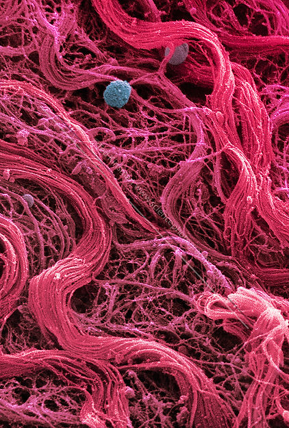
Scanning electron microscope (SEM) image of human muscle tissue collected from an 18 year old
Magnified Image Photograph - Coloured Sem Of Skeletal (striated) Muscle Fibres by Steve Gschmeissner